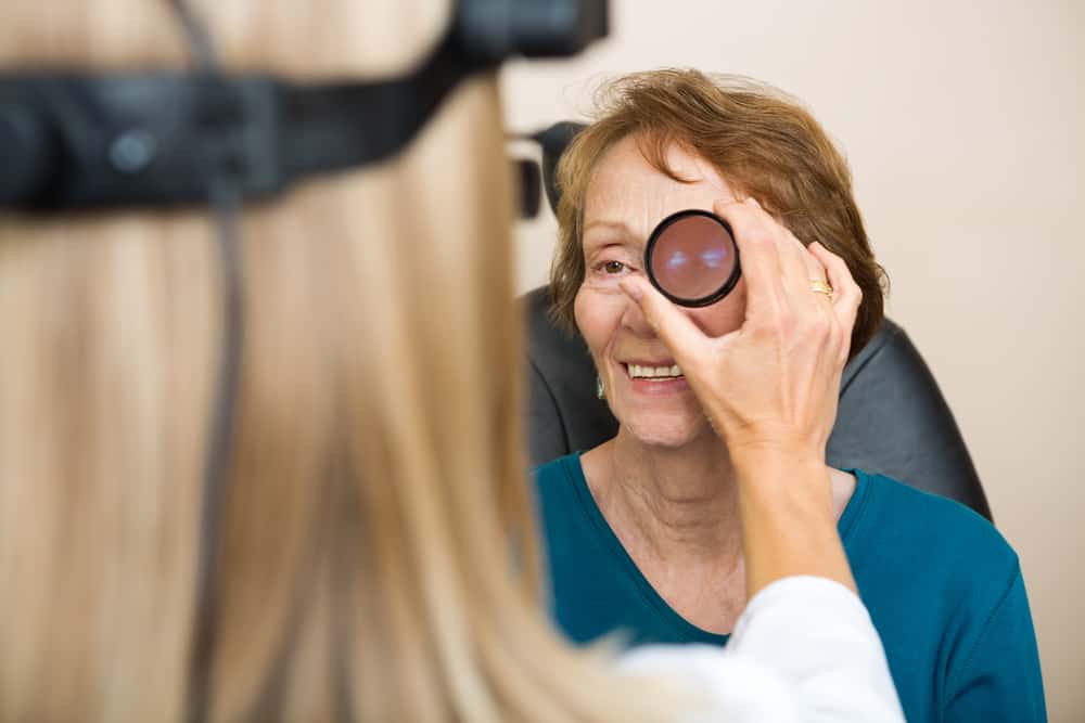
Your Initial Visit
Your initial visit will last approximately 1.5 to 3 hours depending on the type of evaluation and the diagnostic testing needed.
Please bring with you to your appointment:
- Completed new patient forms
- Your insurance cards
- Copays for specialists as noted on your insurance card
- Any referral forms from your referring or primary care physician
- A list of all your medications and eyedrops you are using
- Any contact lenses or glasses that you currently wear for everyday use.
On the day of your visit, your eyes will be dilated (eye drops will be placed to open your pupils). This dilation typically lasts 4-6 hours; therefore, we recommend that you are accompanied by a driver. Your retina specialist will perform a complete evaluation and comprehensive eye exam. Your diagnosis will then be discussed and questions answered. In addition to the eye exam, specific tests may be ordered to help better diagnose and treat your retinal condition. These tests include:
Digital Fundus Photography – A special camera will be used to obtain color photographs of your retina. This is a non-invasive test. These photographs are often obtained to document findings in your retina and used for future comparison.
Fluorescein Angiography – This is a diagnostic test that involves taking photographs of the blood vessels and blood flow in the retina. It involves the use of fluorescein dye, a vegetable-based dye that is injected through a peripheral vein in your arm or hand. After injection of the dye, a special camera is use to capture a series of images of the dye as it circulates through your retina. These images are helpful in diagnosing specific retinal diseases, to assist in determining specific treatments, and to monitor disease progression and treatment response. Please note, this dye is not the same dye used in CT or MRI scans. Fluorescein dye is considered safe for the vast majority of patients and does not contain iodine. Since it is unrelated to other common intravenous dyes, it can be used in patients who have had a reaction to other contrast dyes used by radiologists and other specialists. Please note that patients who receive a fluorescein injection can expect their urine to be a bright yellow-orange color for several hours after the test.
Optical Coherence Tomography (OCT) – This is an advanced, non-invasive technique where a specialized camera captures light rays used to create a cross-sectional image of your retina. These images can help with the detection, treatment, and monitoring of many retinal conditions, such as macular holes, macular puckers, retinal swelling from diabetic retinopathy or macular degeneration. Unlike other imaging techniques that use radiation or sound waves, OCT uses light to produce high resolution images. Therefore, there is no risk of side effects to the eye.
Fundus Autofluorescence (FAF) – This is a non-invasive imaging technique that can evaluate the health of the retina without contrast dye. A camera captures a response from a specific molecule underneath the retina called lipofuscin. This molecule is a fluorophore because it has fluorescent properties when exposed to a specific wavelength of light. Lipofuscin accumulates naturally with age, while its excessive accumulation or absence can be an indicator of specific disease processes. Autofluorescence imaging uses these properties to record the natural fluorescence of the back of the eye to help diagnose and monitor certain diseases, such as macular degeneration, hereditary retinal degenerations, inflammatory diseases, and toxicity from the long term use of a medications, such as plaquenil. Since fundus autofluorescence uses light to produce images, there is no risk of side effects associated with this technique.
B-Scan Ultrasonography – Ultrasonography is a diagnostic technique that uses sound waves to capture of image of the back of the eye. It is similar to the machine used for expectant mothers to image the developing baby. It is usually used in patients who have a dense cataract or severe bleeding inside the eye, which prevents the doctor from seeing into the back of the eye. The procedure can help detect retinal detachments, hemorrhages, inflammation, and tumors in the back of the eye when they cannot be normally viewed with a dilated eye exam. The images obtained of your retina and vitreous can ultimately provide your doctor with important information for diagnosis and treatment planning. Ultrasonography is a painless, non-invasive technique performed in the office. It is safe for all patients and does not involve any radiation exposure.
Optical Coherence Tomography (OCT) – This is a fast, non-invasive technique where camera captures special light rays used to create a cross-sectional image of your retina. These images can reveal areas of abnormalities in your retina. The images can be used to monitor disease progression and treatment response.
Fundus Autofluorescence (FAF) – This is a new, non-invasive imaging technique that uses a special camera to captures a response from specific molecules underneath the retina to show areas of affected or damaged retina. These images can be used to monitor disease progression over time.
B-Scan Ultrasonography – This technique uses an ultrasound machine, similar to the machine used for expectant mothers to image the developing baby, to visualize the back of your eye. The images obtained of your retina and vitreous can provide your doctor with important information for diagnosis and treatment planning.
Your retina specialist will then come up with a plan of treatment. He will then discuss the treatment options with you and your family. The examination and these ancillary tests do take up time. We do appreciate your patience.

